


Groundbreaking Whole-Brain Imaging Reveals Glioma Heterogeneity and Vascular Disruption
In a recent study published in Science Advances, a joint team from Lingang Laboratory and the Shanghai Institute of Materia Medica (SIMM) of Chinese Academy of Sciences (CAS) has constructed the high-resolution, whole-brain panorama of glioma vasculature, uncovering critical details about tumor heterogeneity, blood-brain barrier (BBB) disruption, and nanoparticle (NP) distribution. The research team leverages advanced micro-optical sectioning tomography (MOST) technology to visualize brain tumors at submicron resolution across multiple stages of development.
Using the MOST system combined with whole-brain Nissl staining and 3D reconstruction, the team mapped pathological features of orthotopic glioma in mice at early, intermediate, and advanced stages. Key findings include:
Unexpected Invasiveness Pattern: Early-stage glioma showed diffuse infiltration along host vessels, migrating up to 680 μm along the middle cerebral artery within just four days post-implantation.
Vascular Remodeling: Tumors progressively hijacked and remodeled host vessels, leading to dilated, disorganized, and leaky vascular networks.
Vascular Mimicry (VM): Advanced tumors formed non-endothelial, channel-like structures that connected to existing vasculature, contributing to blood supply.
BBB Permeability: Large-diameter vessels—not just capillaries—became major sites of NP extravasation as the tumor progressed.
Nanoparticles distribution: The distribution of angiopep-2-functionalized PLGA nanoparticles (PLGA-A2), a promising drug delivery system, was also visualized. The NPs preferentially accumulated near leaky large vessels, with penetration depths averaging 12.19 μm. This challenges the conventional view that only capillaries facilitate drug delivery into tumors.
The research bridges the gap between cellular-level detail and whole-organ context, and the multiscale atlas not only deepens our understanding of glioma biology but also provides a practical framework for designing more effective nanomedicines. The research highlights the potential of MOST/fMOST systems in pathological studies and therapeutic development. By revealing how tumor vasculature evolves and influences drug distribution, the study offers a foundation for future strategies in vascular normalization, anti-co-option therapy, and personalized medicine.
DOI: 10.1126/sciadv.adw8330.
Link: https://www.science.org/doi/10.1126/sciadv.adw8330
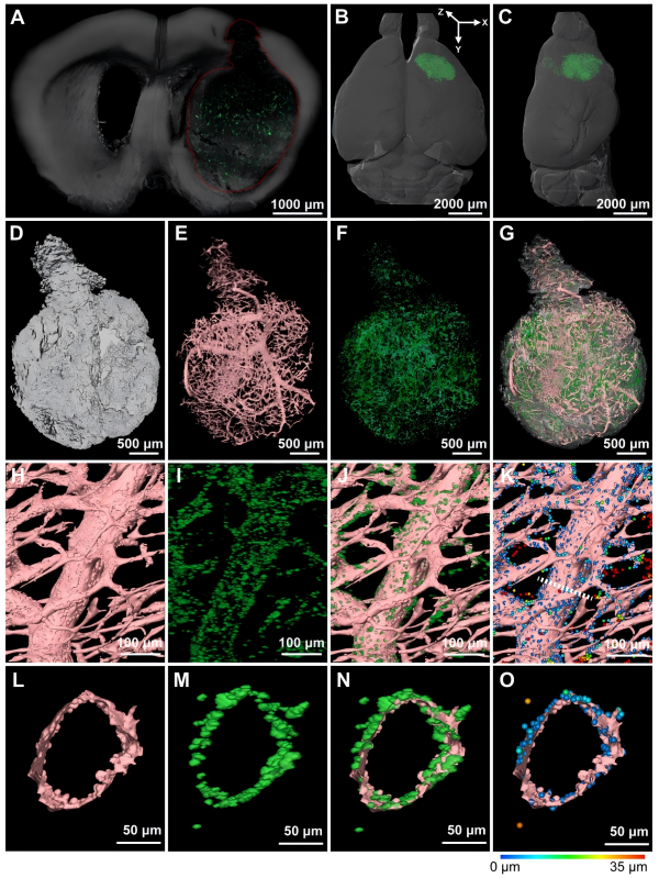
3D reconstruction of glioma vasculature (green) and nanoparticles (yellow) in a whole mouse brain, demonstrating spatial heterogeneity and perivascular NP accumulation. (Image by XIN Xiaohong)
Keywords:
Glioma heterogeneity, whole-brain imaging, nanoparticle distribution
Contact:
DIAO Wentong
Shanghai Institute of Materia Medica
E-mail: diaowentong@simm.ac.cn