


Researchers Highlight Non-invasive Design Strategy Underlying Organ Disease Biomarkers
Non-invasive methods for real-time observation of organ-specific disease biomarkers will help study pathological changes in specific organs and facilitate early diagnosis and effective treatments of diseases.
Fluorescent bioimaging-based assays are widely used in disease biomarker detection due to their high sensitivity, operation simplicity, fast response, high spatio-temporal resolution and non-destructiveness in situ imaging in vivo.
Recently, in a tutorial review published in Chemical Society Reviews, a team of researchers led by Li Jia at the Shanghai Institute of Materia Medica of the Chinese Academy of Sciences, collaborating with research groups led by HE Xiaopeng from East China University of Science and Technology, by Tony D. James from the University of Bath and by Jonathan L. Sessler at the University of Texas at Austin, highlighted the design strategy that underlies the preparation of various promising probes, their optical response to key biomarkers, and proof-of-concept biological studies.
The tutorial review will inspire further study of small-molecule fluorescent probes and pathogenic conditions that contribute to organ-related diseases.
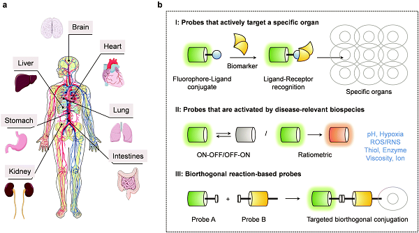
schematic diagram of a) the major organs in the human body considered in the context of the present review and b) the three main design strategies that have been used to create small molecule-based fluorescent probes capable of tracking major organ disease. (image by LI Jia’s laboratory)
Contact:
DIAO WENTONG
Shanghai Institute of Materia Medica of the Chinese Academy of Sciences
E-mail: diaowentong@simm.ac.cn
Reference
Small-molecule fluorescence-based probes for interrogating major organ diseases