Researchers Develop Tracheal Targeted Nanogrid Drug Delivery Systems Visualized by Three-dimensional Pathological Mapping
Understanding how drug delivery systems distribute in vivo remains a major challenge in developing nanomedicines. Especially in the lung, its complex and dynamic microenvironment often limits the effectiveness of existing approaches. Traditional two-dimensional (2D) techniques fail to capture the full spatial and cellular context of how nanocarriers behave in such environments. To address this challenge, “structural pharmaceutics” has been introduced as a new strategy to connect nanoparticle structures with physiological structures through advanced Three-dimensional (3D) imaging and cross-scale characterizations.
In a study published in ACS Nano, a research team led by YIN Xianzhen from the Lingang Laboratory and ZHANG Jiwen from the Shanghai Institute of Materia Medica (SIMM) of the Chinese Academy of Sciences (CAS), reported the development of a precise targeting strategy for tracheal inflammation using nanogrid-based delivery systems composed of gridded cyclodextrin cross-links (GCC). Micro-optical sectioning tomography (MOST) and fluorescence MOST (fMOST) systems enabled multiscale 3D visualization and in-depth pharmacodynamic evaluation.
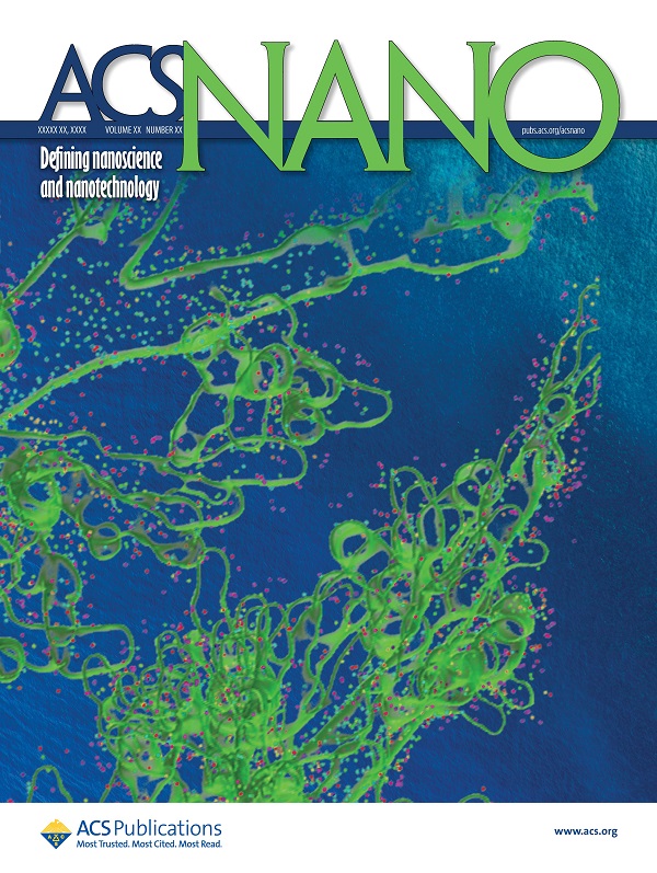
The researchers designed GCC carrier using crosslinked cyclodextrins with effective reactive oxygen species (ROS) scavenging abilities. They then labeled the nanogrid with Rhodamine 110 (Rh110) and tracked its journey after tail vein injection in mice using single-particle tracing and 3D whole-lung imaging. Surprisingly, the nanogrid showed significant accumulation along the outer wall of the trachea, a feature rarely observed with conventional nanoparticles.
When loaded with dexamethasone (DEX), a commonly used corticosteroid, the GCC system not only prolonged drug retention in vivo but also displayed a controlled-release profile. The formulation also showed strong anti-inflammatory effects in a lipopolysaccharide (LPS)-induced bronchitis mouse model. Mice treated with DEX@GCC recovered body weight more rapidly, exhibited improved lung function, and showed significantly reduced inflammatory markers in bronchoalveolar lavage fluid compared to free DEX (higher dose) or model groups.
To visualize these therapeutic outcomes, the researchers developed a whole-lung 3D pathological atlas. Combining fluorescence imaging and machine learning-assisted cell recognition, they reconstructed spatial maps of inflammation and quantified structural changes such as tracheal wall thickening. Virtual endoscopic imaging allowed them to “see” the inside of diseased tracheas and quantitatively assess effects in a non-invasive yet highly detailed manner. These techniques helped confirm the structural repair and anti-inflammatory efficacy of DEX@GCC at both tissue and cellular levels.
This study marks a key step in bridging material science and biomedical imaging. By integrating the concept of "structural pharmaceutics" with 3D organ-level mapping, the team provided a new framework for understanding how nano-drugs behave in complex biological environments. The method opens new possibilities for precise design and evaluation of nanomedicines, especially for pulmonary diseases with high spatial heterogeneity such as lung tumors, bronchitis, and lung fibrosis. This work provides a multiscale lung atlas that bridges basic research and clinical translation. 3D pathology revealed spatiotemporal patterns of progressive lesions to support phenotype screening, target discovery and intervention design.
DOI: 10.1021/acsnano.5c06694
Article link: https://doi.org/10.1021/acsnano.5c06694

Nanogrid-targeting coral-inspired airway models with GCC particle accumulation for enhanced drug delivery (Image by CAO Zeying)
Contact:
DIAO Wentong
Shanghai Institute of Materia Medica, Chinese Academy of Sciences
E-mail: diaowentong@simm.ac.cn


